Weight of The Uterus: A Detailed Look at Uterine Dimensions and Mass

The uterus is a remarkable, dynamic structure within the female reproductive system, playing a pivotal role in menstruation, pregnancy, and childbirth. Although many focus on its function, understanding its physical properties—particularly its heft—can offer deeper insight into how it adapts to a person’s changing life phases. This discussion delves into the background and biology surrounding the female womb, the normal range of its mass, and the numerous factors that can influence the weight of the uterus. By clarifying developmental stages, reproductive transitions, and common medical conditions, this article aims to provide a comprehensive perspective on uterine mass. The information presented here is grounded in current medical research and seeks to highlight key considerations for anyone curious about this essential organ.
A Brief Overview of Uterine Structure
Before exploring why this organ’s heaviness shifts, it helps to gain some clarity on its basic anatomy. The uterus resides in the pelvis, between the bladder and the rectum, and features distinct regions:
- Fundus: The top portion, curved and often the site where the fallopian tubes connect.
- Body (Corpus): The central region that expands during pregnancy and contracts during labor.
- Cervix: The lower segment, connecting the uterus to the vaginal canal.
Composed mostly of muscular tissue (the myometrium) and lined internally with endometrium, this organ has extraordinary capabilities. Its contractile force can push an infant into the world, yet it must remain soft and accommodating for fetal development over many months [1].

Typical Measurements and General Facts
Every uterus is unique, shaped by a person’s genetics, hormonal milieu, and reproductive history. Though commonly described as pear-shaped, its dimensions can vary. For a broader perspective on uterine size variations, understanding the normal size uterus before and after pregnancy provides valuable insight into how this organ adapts throughout different life stages. In an adult who has never been pregnant, the organ often measures around three inches long and about two inches wide. After one or more pregnancies, it may be slightly larger. But size alone doesn’t tell the entire story.
For those wondering how much does uterus weigh, a typical non-pregnant uterus often falls around 50 to 70 grams (roughly 1.8 to 2.5 ounces). In some individuals, it can be marginally lighter or heavier without indicating any pathology. This weight largely reflects the amount of muscle and supportive tissue, as well as blood flow to the region [2].
Influences of Hormones on Uterine Mass
The ovaries produce estrogen and progesterone—two hormones central to reproductive function. Their levels shift during menstrual cycles and across different life stages. Consequently, their influence on uterine size and weight is also considerable.
- Menstrual Cycle
- Endometrial Growth: During the first half of the cycle, estrogen rises, prompting the endometrium to thicken in preparation for potential implantation of a fertilized egg. This buildup can slightly augment uterine weight.
- Shedding: If fertilization does not occur, hormone levels drop and the endometrium sheds, contributing to menstrual bleeding. During this shedding phase, the uterus effectively becomes lighter.
- Pregnancy
- Drastic Growth: When a pregnancy is established, hormonal surges bring about massive expansion. Over the next nine months, the uterus extends far beyond its resting size.
- Weight Increase: By the later stages of pregnancy, the organ can weigh nearly 1 kilogram (2.2 pounds) or more, accommodating the fetus, placenta, and amniotic fluid.
- Menopause
- Decline in Estrogen: As ovarian hormone production decreases, the uterus may shrink or atrophy, often leading to a reduction in mass.
These fluctuations demonstrate how dynamic uterine tissue can be, constantly adapting to the body’s evolving reproductive demands. The cyclical nature of hormonal shifts is key to understanding how the weight uterus can swing over relatively short periods.
Common Conditions That May Affect Uterine Heft
While normal hormonal cycles account for predictable weight changes, certain medical concerns can also alter uterine mass. Below are a few notable examples:
- Fibroids (Leiomyomas)
- Benign Tumors: These growths develop from muscular tissue. They’re common in people of reproductive age and can vary widely in size and number.
- Significant Weight Gain: A uterus dotted with large fibroids might weigh multiple times more than average. In extreme cases, fibroids alone can add several pounds.
- Adenomyosis
- Invasion of Endometrial Tissue: When the lining grows into the uterine muscle, it can thicken the walls and inflate overall mass.
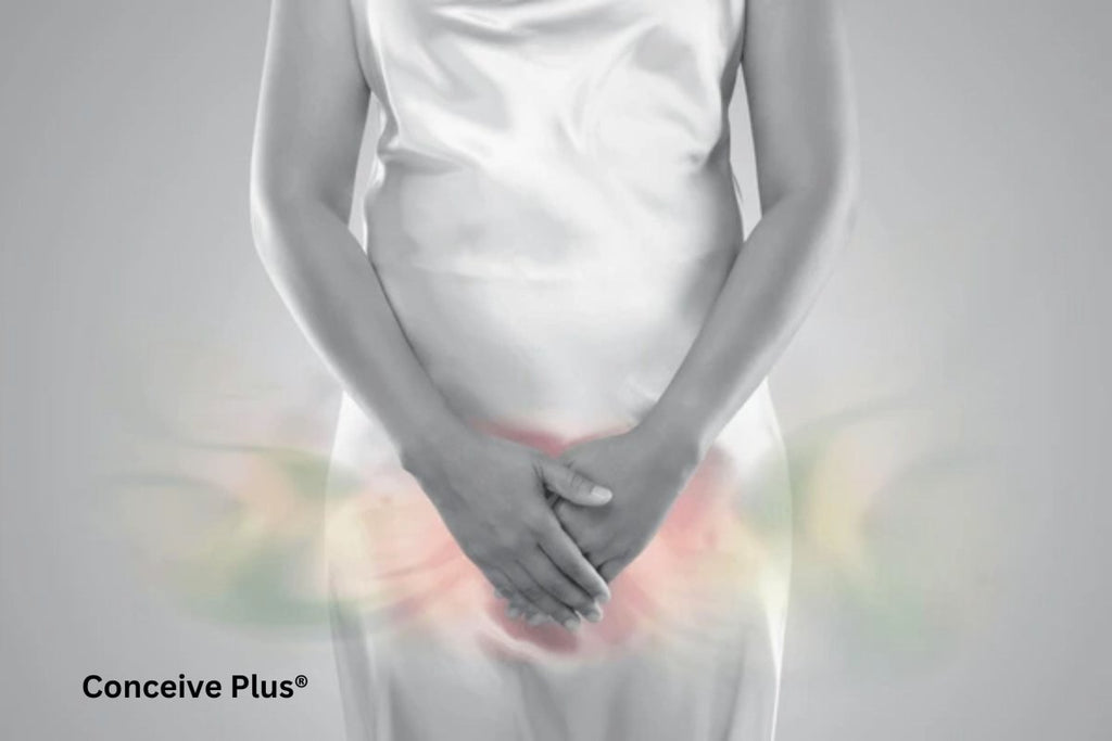
- Symptoms: Many experience heavy menstrual bleeding, pelvic pain, and bloating. This condition can be confirmed through imaging or surgical evaluation.
- Malignancies
- Uterine Cancer: Cancer of the endometrium or other parts of the uterus can introduce abnormal growth and increase its overall mass. Early detection often hinges on symptoms such as unusual bleeding.

✨ Think You Might Be Pregnant?
Get fast, accurate results up to 6 days before your missed period with our early detection pregnancy test.
Shop Now — $9.80✓ Free shipping on orders over $40 | ✓ Trusted by millions of couples worldwide
- Infections or Inflammatory Diseases
- Pelvic Inflammatory Disease: A severe infection affecting the uterus, tubes, and other pelvic structures might lead to swelling or fluid accumulation. This can produce subtle weight changes, though they are typically overshadowed by pain or fever.

Effects of Pregnancy on Uterine Mass
It’s worth devoting extra attention to pregnancy because it brings about some of the most dramatic uterine changes:
- Rapid Early Growth: In the first trimester, hormonal surges contribute to thickening of the lining and initial enlargement of the muscular tissue. By around 12 weeks, the uterus is often large enough to extend above the pubic bone.
- Mid-to-Late Pregnancy: As the fetus grows, the uterus stretches in all directions. Toward the final trimester, it can reach up to the rib cage, carrying not just the baby but the placenta and amniotic fluid.
- After Childbirth: This organ undergoes a process called involution, a natural return toward its pre-pregnancy size. Within six weeks postpartum, it contracts significantly, though it may not fully revert to its exact original dimensions or mass.
The upward weight shift is dramatic. Some studies note the womb may weigh nearly 1 to 2 pounds by the end of gestation, not accounting for the occupant(s). This scenario clarifies how much does uterus weigh under maximum load. The transformation is astonishing for first-time mothers and even more pronounced for those carrying multiple babies (twins, triplets, or higher-order multiples). The body’s capacity to expand, sustain, and then shrink back remains one of nature’s most impressive feats.
Dietary and Lifestyle Factors
While hormone levels and reproductive events dominate the conversation about uterine mass, diet and lifestyle also offer subtler influences:
- Body Weight and Composition: Extreme weight fluctuations can sometimes affect hormonal balance, though the impact on uterine tissue is often indirect. Obesity, for instance, can alter estrogen levels produced by fat cells, potentially influencing uterine growth over the long term.
- Physical Activity: Regular exercise promotes optimal circulation. Some research suggests good blood flow can keep reproductive organs, including the womb, in a healthier state, although it may not drastically alter weight of a uterus in isolation.
- Nutrient Intake: Iron, calcium, and other micronutrients are critical to general reproductive health. A well-rounded diet supports robust blood supply and muscular function of the uterus.
- Smoking and Alcohol: Both can negatively affect fertility and hormone regulation. While direct effects on uterine weight are not commonly cited, unhealthy habits may create an environment more prone to pathological changes [3].
Medical Imaging and Diagnostic Methods
When clinicians need to ascertain the weight uterus or gauge its volume and structure, they rely on modern imaging modalities. These tools are crucial for identifying abnormalities, measuring changes, and planning treatments:
- Ultrasound
- Transabdominal and Transvaginal: Both approaches generate images of the womb, providing insights on thickness, fibroids, and other growths.
- Doppler Ultrasound: Evaluates blood flow, helping detect vascular malformations or suspicious masses [4].
- Magnetic Resonance Imaging (MRI)
- High-Resolution Detail: MRI offers sharper contrasts of soft tissues, making it useful for complex cases, such as deeply embedded fibroids or adenomyosis.
- Computed Tomography (CT) Scan
- Less Common for Uterus: CT scans can be used, though radiation exposure and reduced soft-tissue differentiation mean they’re not typically the first choice.
By combining patient history, physical exams, and these imaging techniques, healthcare professionals can accurately estimate the weight of a uterus. This data informs key decisions, from monitoring fibroids to planning any necessary surgical approach.
The Emotional Dimension: Empowerment Through Knowledge
For some, the idea of investigating the weight uterus may seem purely clinical. Yet for many people, it’s part of a broader journey of learning about bodily processes—an empowering step toward making informed decisions about healthcare and lifestyle. Individuals who have endured gynecological conditions often articulate that understanding the biological underpinnings provides a sense of agency, lessening anxiety about what might be happening internally.
- Body Positivity: Embracing the body’s natural fluctuations can help demystify changes encountered during menstruation, pregnancy, or menopause.
- Informed Advocacy: Knowing typical ranges and red flags better equips patients to communicate concerns or request tests when something feels amiss.
- Shared Experience: Many find solace in discussing these topics within support groups or online forums. As more people openly talk about experiences like fibroid removal or postpartum healing, the sense of isolation diminishes.

The Bottom Line
With all of this in mind, it is clear that understanding the weight of the uterus is not merely an exercise in academic curiosity, but an essential component of recognizing how profoundly the womb interacts with various aspects of human health.
Ultimately, uterine weight fluctuates based on a myriad of factors—ranging from monthly hormone cycles and pregnancy to medical conditions like fibroids. Answering “how much does uterus weigh?” requires context: a typical, non-pregnant adult womb might be 50-70 grams, whereas during pregnancy, it grows substantially to accommodate fetal development. Conditions such as fibroids, adenomyosis, or cancer can also alter uterine heft and warrant focused attention if symptoms arise.
Given the enormous breadth of normal variation and the organ’s capacity for change, the question “what is the weight of a uterus?” cannot be answered with a single fixed number. Rather, it represents a starting point for deeper investigation into reproductive health. For anyone curious about their own uterine status, speaking with a healthcare provider and, if needed, undergoing imaging helps clarify where one sits on the spectrum. Knowledge is a cornerstone of informed decision-making, and the more individuals understand about their reproductive anatomy, the better equipped they are to navigate each phase of life.
References
- Ameer, M. A., Fagan, S. E., Sosa-Stanley, J. N., & Peterson, D. C. (2022). Anatomy, Abdomen and Pelvis: Uterus. In StatPearls. StatPearls Publishing. Available at: https://pubmed.ncbi.nlm.nih.gov/29262069/
- Gao, H., Liu, D. E., Li, Y., Tang, J., Hu, S., Wu, X., Tian, Z., & Tan, H. (2019). Uterine size and volume are associated with a higher clinical pregnancy rate in patients undergoing assisted reproduction technology: A longitudinal study (A STROBE-compliant article). Medicine. Available at: https://pubmed.ncbi.nlm.nih.gov/30813136/
- Rostand, A., Kaminski, M., Lelong, N., Dehaene, P., Delestret, I., Klein-Bertrand, C., Querleu, D., & Crepin, G. (1990). Alcohol use in pregnancy, craniofacial features, and fetal growth. Journal of epidemiology and community health. Available at: https://pubmed.ncbi.nlm.nih.gov/2277252/
- Ulrich, C. C., & Dewald, O. (2023). Pregnancy Ultrasound Evaluation. In StatPearls. StatPearls Publishing. Available at: https://pubmed.ncbi.nlm.nih.gov/32491504/
🌱 Get Answers Early
Millions of women trust Conceive Plus for their fertility journey. Clinically validated pregnancy tests delivered to your door.
Shop Pregnancy Tests →Frequently Asked Questions
Recommended by Fertility Experts
Conceive Plus Men's Fertility Support
Designed to support healthy sperm production, motility, and overall male reproductive health with clinically studied nutrients.
Shop Now →Q: How much does a womb weigh?
A: A non-pregnant uterus typically weighs between 30 to 40 grams (approximately 1 to 1.4 ounces) in women of reproductive age, though this can vary based on factors like age, hormonal status, and reproductive history. The uterus is a relatively small, pear-shaped organ measuring about 7 to 8 centimeters in length and 4 to 5 centimeters in width when not pregnant. During pregnancy, the uterus expands dramatically and can weigh up to 1,000 grams (2.2 pounds) by the third trimester as it accommodates fetal development. After menopause, the uterus typically shrinks and may weigh even less due to decreased estrogen levels and tissue atrophy. Understanding normal uterine dimensions helps healthcare providers identify potential abnormalities during routine gynecological examinations.
Q: What affects uterine weight and size?
A: Several factors influence uterine weight and dimensions, including age, hormonal fluctuations, pregnancy history, and overall reproductive health status. Women who have had multiple pregnancies often have slightly larger and heavier uteri compared to nulliparous women, as the uterine muscle tissue (myometrium) undergoes permanent remodeling. Estrogen levels play a crucial role—younger women of reproductive age typically have heavier uteri than postmenopausal women due to higher circulating estrogen. Medical conditions such as uterine fibroids, adenomyosis, and polycystic ovary syndrome (PCOS) can also increase uterine weight by causing tissue growth and inflammation. Maintaining overall reproductive health through proper nutrition, stress management, and supplementation with evidence-based fertility support like Conceive Plus can help optimize reproductive system function during your fertility journey.
Q: Does uterine weight matter for fertility?
A: While uterine weight itself isn't a direct fertility marker, the underlying conditions that affect it—such as fibroids, adenomyosis, or inflammation—can significantly impact fertility and pregnancy outcomes according to the American Society for Reproductive Medicine (ASRM). A healthy, normally-weighted uterus with good blood flow and minimal inflammation provides the optimal environment for embryo implantation and fetal development. Abnormal uterine enlargement may indicate conditions that reduce fertility rates by 30-40%, making proper diagnosis and management important for those trying to conceive. Regular pelvic ultrasounds can assess uterine dimensions and rule out pathological changes that might affect conception or pregnancy success. Supporting your reproductive health with comprehensive wellness strategies, including prenatal supplementation formulated to support fertility, is an important part of optimizing your chances of conception.
Q: What's the difference between uterine weight before and after pregnancy?
A: The uterus undergoes dramatic changes during pregnancy—expanding from its typical 30-40 gram weight to approximately 1,000 grams at term, representing a roughly 25-fold increase in mass. This expansion is driven by both myometrial muscle growth and the accumulation of amniotic fluid, placental tissue, and the developing fetus, which collectively account for several pounds of pregnancy weight. After delivery, the uterus begins a process called involution, where it rapidly shrinks back toward its pre-pregnancy size and weight over a period of 6 to 8 weeks postpartum. However, women who have completed pregnancies may retain a slightly heavier uterus long-term compared to nulliparous women, as some myometrial changes persist permanently. Understanding these changes helps women appreciate the remarkable adaptability of their reproductive system throughout different life stages.
Q: Can uterine weight be measured during gynecological exams?
A: While gynecologists cannot precisely measure uterine weight during a standard clinical examination, they can assess uterine size and approximate mass through bimanual pelvic exams and transvaginal ultrasound, which provides detailed dimensional measurements. Transvaginal ultrasound is the gold standard for evaluating uterine dimensions and can detect abnormal enlargement that may indicate conditions like fibroids or adenomyosis affecting reproductive health. The American College of Obstetricians and Gynecologists (ACOG) recommends routine pelvic ultrasounds as part of comprehensive fertility evaluations to assess uterine health and rule out structural abnormalities. Precise weight calculations can be performed using 3D ultrasound technology, which calculates volume and estimates mass based on standardized formulas used in research and clinical settings. Regular monitoring of uterine health through appropriate imaging is an important component of preconception care for those planning to conceive.
Trusted by Couples in Over 70 Countries
Support Male Fertility From the Inside Out
Male fertility is just as important as female fertility in the conception journey. Conceive Plus Men's range delivers targeted nutritional support to promote healthy sperm and overall reproductive wellbeing.
Shop Men's Range →