Bladder Uterus Anatomy Differences In Males and Females
There are many differences in male and female anatomy, and understanding those differences can help you better understand female body functions. The bladder uterus anatomy in males and females is an example of these differences. The bladder is slightly different in shape and orientation in women compared to men [1]. The uterus is only present in women and is the vital organ of female fertility [2].
In this article, we will explore the structure, function, and differences between uterus and bladder anatomy in males and females. This article also includes the role of uterus bladder in reproductive health or fertility.

The Female Bladder
The female bladder is a hollow, muscular organ located in the pelvis, just behind the pubic bone and in front of the uterus. When the bladder is empty, it appears as an inverted triangle, and when it stores urine, it expands to fill. The bladder wall has three main layers:
- Inner layer: The innermost layer is called urothelium, and it prevents urine from leaking into surrounding tissues.
- Middle Layer: The middle layer is called the detrusor muscle, and this muscle helps the bladder contract to expel urine.
- Outer Layer: The outermost layer is of connective tissue that provides support.

The bladder connects to the outside of the body through the urethra. In females, the urethra is much shorter than in males, measuring about 1.5 inches or 4 cm in length [3]. This shorter urethra is a major reason why women are more likely to experience urinary tract infections (UTIs) more often [4].
Function of the Bladder
The primary function of the bladder is to store and expel urine. Kidneys produce urine and transfer it to the bladder through tubes called ureters. Once the bladder is full, nerves signal the brain, creating the urge to urinate.
During urination, the middle layer of the bladder or detrusor muscle contracts, and the urethral sphincter — two muscles that regulate the opening of the urethra — relaxes to allow urine to pass out of the body.
Differences from Male Anatomy
The female bladder differs from the male bladder mainly in its position in the women pelvis and the length of the urethra. In males, the bladder is located above the prostate gland and has a longer urethra. This longer urethra makes it harder for germs to reach inside the urinary tract and cause UTIs. This is why UTIs are less common in men compared to women.
The Female Uterus
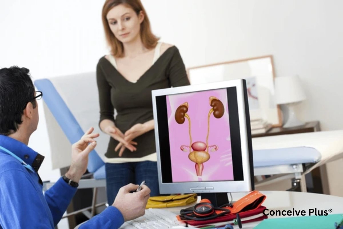
The uterus is among the primary reproductive organs in females. It is a pear-shaped, muscular organ located in the female pelvis. Its location is behind the bladder and in front of the rectum [2].
The uterus has three main parts:
- Fundus — The upper, rounded part.
- Body — The main part where a fertilized egg implants during pregnancy.
- Cervix — The lower part that opens into the vagina.

Like the bladder, the uterine wall also has three layers, which are:
- Endometrium: Enometrium is the inner lining of the uterus and has a very important role in female reproduction and fertility health [5]. It is the layer that thickens during the menstrual cycle to prepare for the potential implant after fertilization. However, if the implant does not occur, this layer sheds and is characterized by menses.
- Myometrium: It is the middle layer of the uterus wall, which is mainly made up of smooth muscles. These muscles contract during labor and menstruation to expel the baby or shaded endometrium outside.
- Perimetrium: It is the outer protective layer that doesn't perform any specific role in reproduction or pregnancy other than protection.
Function of the Uterus
The uterus plays a central role in female reproduction. It allows the follow of sperm towards the fallopian tube for fertilization, provides space for the implantation of a fertilized egg, and supports the development of a fetus during pregnancy [2].
During labor or when the delivery time approaches, the myometrium layer of the uterus contracts to help deliver the baby. Female fertility hormones like estrogen and progesterone regulate the function of the uterus, whether it be preparing for implantation or nourishing the fetus during pregnancy [6]. For women looking to enhance their fertility, products like Conceive Plus Women's Fertility Support can provide essential nutrients and support to optimize reproductive health.
Differences from Male Anatomy
Males do not have a uterus and it is unique to female anatomy. Instead, the reproductive system in males includes organs like the testes, prostate gland and seminal vesicles. In females, the uterus is essential for reproduction, while the testes, prostate, and seminal vesicles contribute to semen production in males.

✨ Support Healthy Sperm
Our Motility Support supplement contains Zinc, CoQ10, and L-Carnitine — clinically studied nutrients for sperm health and motility.
Shop Now — $34.95✓ Free shipping on orders over $40 | ✓ Trusted by millions of couples worldwide

Interplay Between Bladder and Uterus
Although the bladder and uterus are both part of different organ systems, they do have some similarities in terms of their anatomical position and nerve and blood supply.
Anatomical Positions
The bladder and uterus are located close to each other in women pelvis, and their positions can affect one another. For example, when the bladder is full, it can press against the uterus, slightly shifting its position. Similarly, during pregnancy, the growing uterus presses on the bladder, reducing its capacity and causing frequent urination. The position of the uterus, whether it is anteverted vs retroverted uterus, can influence its relationship with the bladder and other pelvic organs, potentially affecting urinary and reproductive health.
Shared Vascular and Nervous Systems
The bladder and uterus share blood supply from nearby arteries, such as the internal iliac artery [7]. They also share a network of nerves that control their functions. This close connection can sometimes lead to conditions that affect both organs, such as pelvic pain or pressure.
Medical Conditions that Affect Bladder
Here are some common medical conditions that target the bladder:
- Urinary Tract Infections: UTIs can affect any part of the urinary system, including the bladder. These infections are common in women because they have a shorter urethra compared to men [4]. Symptoms of UTIs can include burning during urination and frequent urges to urinate.
- Overactive Bladder: This condition causes a frequent and sudden urge to urinate, often without much warning. It occurs when the bladder muscles start to tighten on their own.
- Urinary Incontinence: Urinary incontinence refers to involuntary leakage of urine. This condition often occurs due to weakened pelvic muscles after childbirth or aging.
Medical Conditions that Affect Uterus
Here are some uterus-related medical conditions:
- Fibroids: These are non-cancerous growths in the uterine wall and surrounding tissues that can cause heavy periods and pain. These non-cancerous growths are mostly made up of muscles and fibrous tissues, and they can make it hard to conceive [8].
- Endometriosis: A condition where tissue similar to the endometrium grows outside the uterus. It causes pain and discomfort and also decreases the chances of getting pregnant.
- Uterine Prolapse: Uterine prolapse happens when the uterus moves down into the vaginal canal. This occurs because the pelvic floor muscles become weak.
Differences in Male And Female Urinary System
The major difference between male and female urinary tract is the size of the urethra. In males, the urethra is about 8 inches or 20 cm long. On the other hand, the female urethra is about 1.5 inches or 4 cm long only. The shorter urethra increases the risk of UTIs since foreign infectious particles can enter through the shorter urethra into the urinary tract.
The bladder location also has some anatomical differences between males and females. In females, the bladder is located in the pelvis, in front of the uterus and above the vagina. However, in males, the bladder is present above the prostate gland and anterior to the rectum.

Differences In Male and Female Reproductive System
The differences between the male and female reproductive system are more prominent than in the urinary system. The reproductive system in males primarily comprises the penis, testes, prostate gland, and seminal vesicles [9]. Testes produce and store sperm cells, while the prostate gland and seminal vesicles add secretions into the sperm to make it ready for fertilization. Penis is the organ that delivers these sperm cells into the female vagina.
The female reproductive system consists of vagina, uterus, ovaries, and fallopian tubes. Ovaries are the place where the female egg matures and is released. The released egg travels to the fallopian tube, where fertilization occurs. After fertilization, the embryo implants into the uterus.
Hormones that regulate fertility in males and females also differ. Female fertility hormones consist of estrogen and progesterone, while males have testosterone as the primary sex hormone.
The Bottom Line
The male and female bodies have some anatomical differences, and uterus and bladder anatomy are a part of those differences. They are essential organs with distinct roles in the urinary and reproductive systems. The bladder uterus anatomy or location in the pelvis allows them to influence each other's function.
The bladder primarily acts as a urine reservoir, while the uterus is important for many reproductive roles, including fertilization, implantation, and delivery. Studying the anatomy of these organs helps you better understand their role in the urinary and reproductive systems. Also, it helps in proper care and prevention of different health conditions related to these organs.
Resources Used
- Abelson, B., Sun, D., Que, L., Nebel, R. A., Baker, D., Popiel, P., Amundsen, C. L., Chai, T., Close, C., DiSanto, M., Fraser, M. O., Kielb, S. J., Kuchel, G., Mueller, E. R., Palmer, M. H., Parker-Autry, C., Wolfe, A. J., & Damaser, M. S. (2018). Sex differences in lower urinary tract biology and physiology. Biology of Sex Differences, 9(1). https://doi.org/10.1186/s13293-018-0204-8
- Ameer, M. A., Fagan, S. E., Sosa-Stanley, J. N., & Peterson, D. C. (2022, December 6). Anatomy, Abdomen and Pelvis: Uterus. StatPearls - NCBI Bookshelf. https://www.ncbi.nlm.nih.gov/books/NBK470297/
- Urethra | SEER Training. (n.d.). https://training.seer.cancer.gov/anatomy/urinary/components/urethra.html
- Czajkowski, K., Broś-Konopielko, M., & Teliga-Czajkowska, J. (2021). Urinary tract infection in women. Przeglad menopauzalny = Menopause review, 20(1), 40–47. https://doi.org/10.5114/pm.2021.105382
- Critchley, H. O. D., Maybin, J. A., Armstrong, G. M., & Williams, A. R. W. (2020). Physiology of the Endometrium and Regulation of Menstruation. Physiological Reviews, 100(3), 1149–1179. https://doi.org/10.1152/physrev.00031.2019
- Rosner, J., Samardzic, T., & Sarao, M. S. (2024c, March 20). Physiology, Female Reproduction. StatPearls - NCBI Bookshelf. https://www.ncbi.nlm.nih.gov/books/NBK537132/
- Selçuk, İ., Yassa, M., Tatar, İ., & Huri, E. (2018). Anatomic structure of the internal iliac artery and its educative dissection for peripartum and pelvic hemorrhage. Turkish journal of obstetrics and gynecology, 15(2), 126–129. https://doi.org/10.4274/tjod.23245
- Barjon, K., & Mikhail, L. N. (2023, August 7). Uterine Leiomyomata. StatPearls - NCBI Bookshelf. https://www.ncbi.nlm.nih.gov/books/NBK546680/
- Professional, C. C. M. (2024j, November 22). Male Reproductive System. Cleveland Clinic. https://my.clevelandclinic.org/health/body/9117-male-reproductive-system
🌱 Boost Male Fertility Naturally
Conceive Plus Men's Motility Support is formulated with the nutrients that matter most for sperm quality, count, and motility.
Shop Men's Fertility →Frequently Asked Questions
Recommended by Fertility Experts
Conceive Plus Men's Fertility Support
Designed to support healthy sperm production, motility, and overall male reproductive health with clinically studied nutrients.
Shop Now →Q: Why is bladder position different in males and females?
A: The bladder's position and shape differ between sexes due to the presence of the uterus in females, which sits directly in front of the bladder and affects its orientation. In women, the bladder is more anterior and compressed, while in men, it maintains a more rounded shape without this anatomical constraint. This difference is a normal variation in human anatomy and doesn't indicate a health problem. Understanding this distinction helps explain why women may experience different urinary patterns, especially during pregnancy when the growing uterus exerts additional pressure on the bladder. The American College of Obstetricians and Gynecologists (ACOG) notes that these anatomical differences are important for healthcare providers when evaluating reproductive and urinary health concerns.
Q: What does the uterus do in female fertility?
A: The uterus is the primary reproductive organ where a fertilized egg implants and develops into a fetus during pregnancy, making it essential for conception and childbearing. According to the American Society for Reproductive Medicine (ASRM), a healthy uterus with adequate uterine lining thickness (typically 8-12mm) is crucial for successful implantation and pregnancy continuation. The uterine muscles contract during menstruation and labor, while the uterine lining (endometrium) thickens and sheds monthly as part of the menstrual cycle. Many couples undergoing fertility treatment focus on uterine health as a key factor in their success rates. Proper nutrition and supplementation with fertility-supporting nutrients can help optimize uterine health, which is why products like Conceive Plus are designed to support overall reproductive wellness for women trying to conceive.
Q: Can bladder problems affect your ability to get pregnant?
A: While bladder problems themselves don't directly cause infertility, untreated urinary tract infections (UTIs) can potentially impact fertility by causing inflammation and affecting reproductive function. Research indicates that approximately 10-15% of couples of reproductive age experience infertility, and while UTIs are not a primary cause, they should be treated promptly to maintain overall reproductive health. Chronic pelvic conditions affecting the bladder may contribute to pelvic inflammation that could indirectly affect fertility, which is why comprehensive reproductive health is important when trying to conceive. If you're experiencing bladder symptoms alongside fertility concerns, it's important to consult with both a urologist and fertility specialist for proper evaluation. Maintaining overall pelvic and urinary health through hydration, proper nutrition, and supplements designed to support reproductive wellness can be part of a comprehensive fertility plan.
Q: How does the uterus change during the menstrual cycle?
A: The uterus undergoes significant changes throughout the menstrual cycle, with the endometrial lining thickening during the follicular and luteal phases in preparation for potential pregnancy. During the first half of the cycle (follicular phase), estrogen causes the uterine lining to proliferate and thicken, reaching its optimal thickness of 8-12mm by ovulation, according to ASRM guidelines. After ovulation, progesterone transforms the endometrium into a secretory lining suitable for embryo implantation, a critical window that typically lasts 12-14 days. If pregnancy doesn't occur, the drop in hormone levels triggers menstruation, during which the uterine lining sheds through uterine contractions. Supporting hormonal balance and uterine health through proper nutrition and supplementation is important for couples trying to conceive, as optimal endometrial development is essential for successful implantation and early pregnancy establishment.
Q: Why do women have urinary urgency more than men?
A: Women experience more frequent urinary urgency than men partly due to anatomical differences, including a shorter urethra and the positioning of the bladder directly below the uterus, which can create pressure on bladder nerves. The female urethra is only about 1.5 inches long compared to 8 inches in men, making it more sensitive to pressure changes and bladder fullness signals. Hormonal fluctuations during the menstrual cycle also affect bladder sensitivity, with many women reporting increased urinary frequency during certain cycle phases. Additionally, pregnancy significantly increases urinary urgency as the growing uterus places direct pressure on the bladder, affecting approximately 80% of pregnant women according to medical literature. Maintaining proper hydration and supporting overall reproductive health through nutrition and fertility supplements like Conceive Plus can help women manage these normal physiological changes while optimizing their fertility health.
Trusted by Couples in Over 70 Countries
Support Male Fertility From the Inside Out
Male fertility is just as important as female fertility in the conception journey. Conceive Plus Men's range delivers targeted nutritional support to promote healthy sperm and overall reproductive wellbeing.
Shop Men's Range →